Tissue microarray (TMA)
Contact: Senior scientist Matthias Kolberg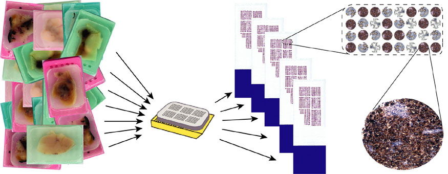
A single TMA block contains of hundreds of individual tumour biopsies and allows for in situ labelling and detection of DNA, RNA, or protein.
Tissue cores from up to several hundreds archival paraffin embedded donor tissue blocks are transferred to a single recipient tissue microarray block as initially described in Kononen et al.1998. Sections from the TMA block are fixed onto microscope slides and can be utilised for immunohistochemical analysis of protein expression in situ. DNA and RNA fluorescence in situ hybridisation (FISH) may also be performed on the TMA slides.
Tumor tissue from the following cancer diseases are studied on TMAs:
- Colorectal cancer (CRC)
- Malignant peripheral nerve sheath tumour (MPNST)
- Ovarian germ cell tumour (OGCT)
- Testicular germ cell tumour (TGCT)
Tumor tissue from the following cancer diseases are studied on TMAs:
- Colorectal cancer (CRC)
- Malignant peripheral nerve sheath tumour (MPNST)
- Ovarian germ cell tumour (OGCT)
- Testicular germ cell tumour (TGCT)